Chinese researchers reveal embryonic mouse brain development with new imaging technique


BEIJING -- Chinese researchers have developed an in vivo embryonic mouse intrauterine imaging technique, which has elucidated the dynamic process of brain development in embryonic mice in vivo, the Science and Technology Daily has reported.
The study, conducted by a research team from Tsinghua University, was recently published in the journal Cell. Experts say this technology holds significant potential for deciphering the mechanisms of developmental disorders.
"The in vivo imaging technique is based on two-photon microscopy and utilizes an auxiliary support device to immobilize the embryonic mouse, enabling long-term, wide-field and deep-tissue live observation," said Mi Da, the corresponding author of the paper.
Mi noted that this technology successfully overcomes the limitations of traditional embryonic in vivo imaging in terms of stability, duration, field of view and operability, allowing researchers to observe various indicators such as embryonic cerebral blood flow and brain tissue cell activity.
Building on this, the research team labeled excitatory and inhibitory neurons in the cerebral cortex of embryonic mice and, combined with intrauterine imaging, comprehensively analyzed the individual and collective migration patterns of different neuron types.
The study provides new in vivo evidence of abnormal neuronal migration in mouse models of neurodevelopmental disorders, while also uncovering the dynamic behavioral characteristics of embryonic immune cells in response to environmental stress.
Shi Songhai, an academician of the Chinese Academy of Sciences, said that the technological framework and analytical methods established in this study will serve as crucial tools for future research on brain development and developmental brain disorders, offering profound methodological significance and broad application prospects.


- Chinese researchers reveal embryonic mouse brain development with new imaging technique
- Taiwan's Lai Ching-te slammed for provocations risking war
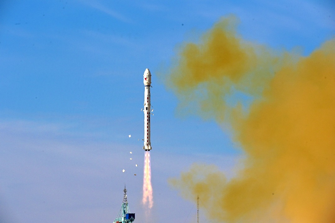
- China launches two new satellites into space
- Technology enhanced weather forecasting accuracy and reduced disaster risks in 2024, report finds
- AS700 airship receives production certificate, readies for flight services
- Xinjiang sees record grain, cotton outputs